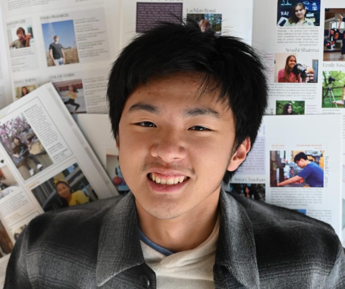


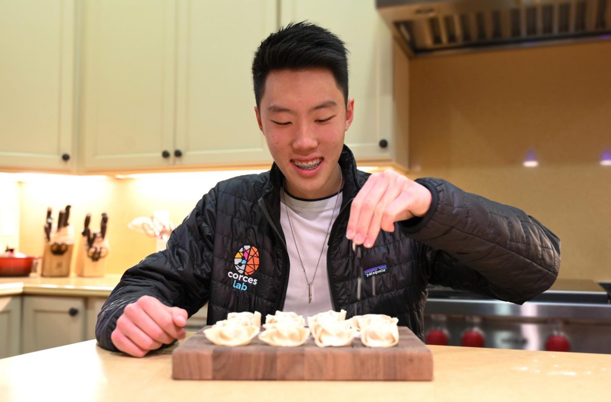
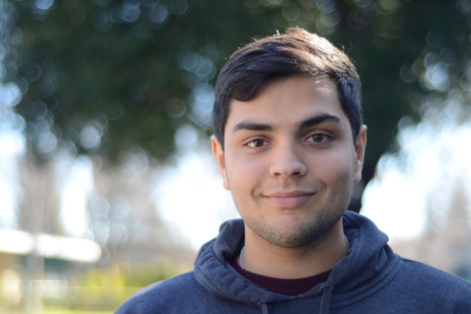
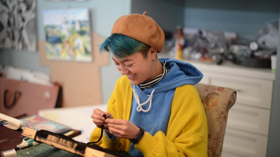
![“[Building nerf blasters] became this outlet of creativity for me that hasn't been matched by anything else. The process [of] making a build complete to your desire is such a painstakingly difficult process, but I've had to learn from [the skills needed from] soldering to proper painting. There's so many different options for everything, if you think about it, it exists. The best part is [that] if it doesn't exist, you can build it yourself," Ishaan Parate said.](https://harkeraquila.com/wp-content/uploads/2022/08/DSC_8149-900x604.jpg)

![“When I came into high school, I was ready to be a follower. But DECA was a game changer for me. It helped me overcome my fear of public speaking, and it's played such a major role in who I've become today. To be able to successfully lead a chapter of 150 students, an officer team and be one of the upperclassmen I once really admired is something I'm [really] proud of,” Anvitha Tummala ('21) said.](https://harkeraquila.com/wp-content/uploads/2021/07/Screen-Shot-2021-07-25-at-9.50.05-AM-900x594.png)

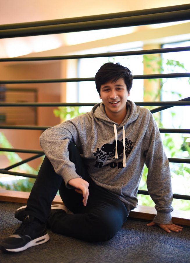


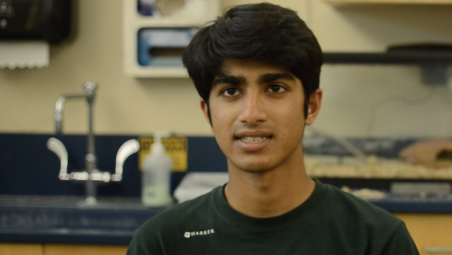
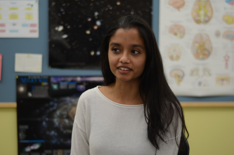
![“I think getting up in the morning and having a sense of purpose [is exciting]. I think without a certain amount of drive, life is kind of obsolete and mundane, and I think having that every single day is what makes each day unique and kind of makes life exciting,” Neymika Jain (12) said.](https://harkeraquila.com/wp-content/uploads/2017/06/Screen-Shot-2017-06-03-at-4.54.16-PM.png)


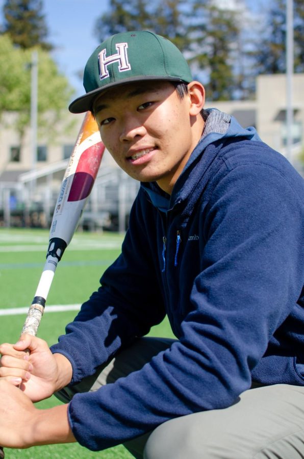
![“My slogan is ‘slow feet, don’t eat, and I’m hungry.’ You need to run fast to get where you are–you aren't going to get those championships if you aren't fast,” Angel Cervantes (12) said. “I want to do well in school on my tests and in track and win championships for my team. I live by that, [and] I can do that anywhere: in the classroom or on the field.”](https://harkeraquila.com/wp-content/uploads/2018/06/DSC5146-900x601.jpg)
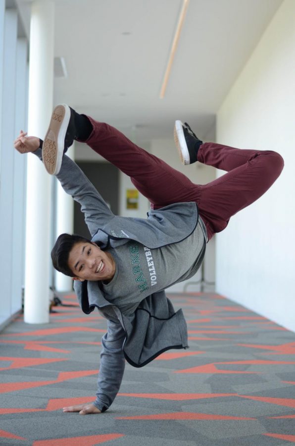
![“[Volleyball has] taught me how to fall correctly, and another thing it taught is that you don’t have to be the best at something to be good at it. If you just hit the ball in a smart way, then it still scores points and you’re good at it. You could be a background player and still make a much bigger impact on the team than you would think,” Anya Gert (’20) said.](https://harkeraquila.com/wp-content/uploads/2020/06/AnnaGert_JinTuan_HoHPhotoEdited-600x900.jpeg)

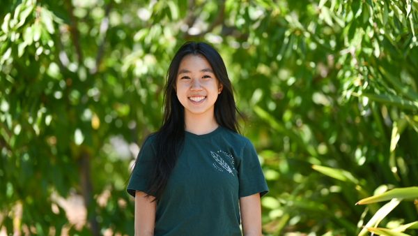
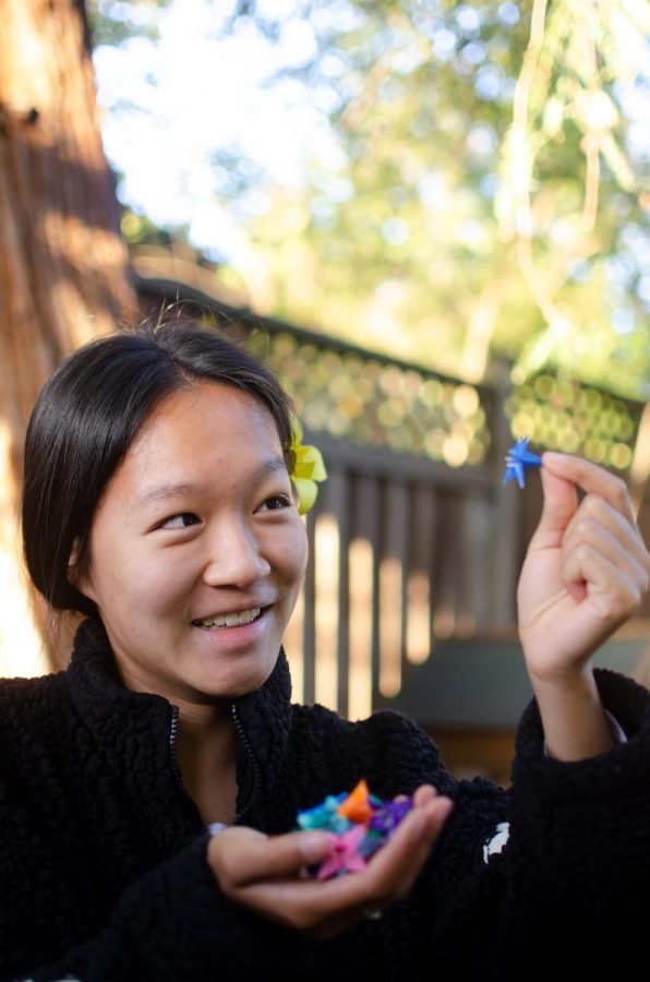
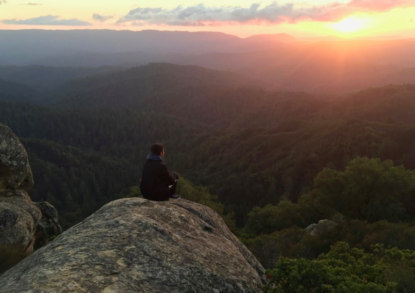
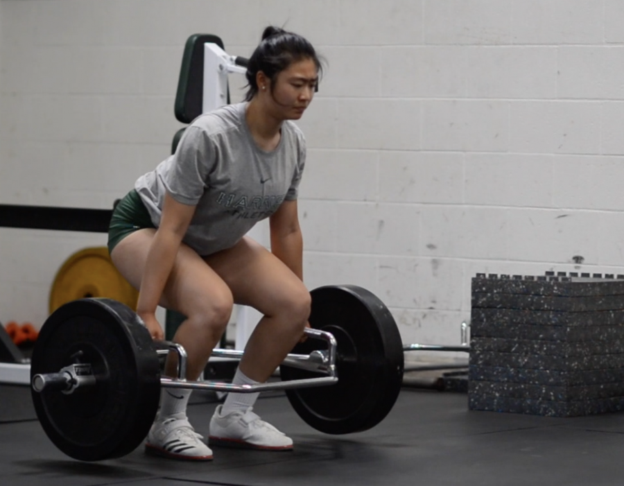
![“I'm not nearly there yet, but [my confidence has] definitely been getting better since I was pretty shy and timid coming into Harker my freshman year. I know that there's a lot of people that are really confident in what they do, and I really admire them. Everyone's so driven and that has really pushed me to kind of try to find my own place in high school and be more confident,” Alyssa Huang (’20) said.](https://harkeraquila.com/wp-content/uploads/2020/06/AlyssaHuang_EmilyChen_HoHPhoto-900x749.jpeg)

This is the eighth installment of Research Revelations: Conversations with Our Student Researchers, a podcast where Aquila staff members talk to student researchers about their projects and research goals. In this episode, News Assistant Editor Chelsea Xie (10) and Features Assistant Editor Elizabeth Zhang (10) meet with Juliana Li (12) to discuss her work in 3D modeling for lung cancer detection.
Chelsea: Hi everyone, I’m Chelsea.
Elizabeth: I’m Elizabeth.
Chelsea: And welcome back to Research Revelations: Conversations With Our Student Researchers.
Elizabeth: Today, we’re with senior Juliana Li to talk about generating a 3D model of branching airway networks for lung cancer detection. So Juliana, thank you so much for joining us. I want to first ask: What was the basis of your research project?
Juliana: So my research project is about lung cancer, and I’m trying to detect and also improve treatment. It’s a bioengineering project through various methodologies of bioengineering: there are bioinformatics, computational anatomical modeling and also AI modeling.
Elizabeth: How did you originally start with this research?
Juliana: I actually started this research because I was placed in a respiratory Dynamics Lab at the University of Iowa, so I applied to that for the bioengineering project. My mentor, the principal investigator of the Hermann Respiratory Dynamics Lab, was really interested in all these respiratory applications of bioengineering, so he suggested to me the project of anatomical modeling. It’s actually a project that he failed to do a couple of years ago. I took on a different perspective to figure out the problem and made an improved model. After that, I did AI work on my own, and then most recently, I’ve been working with a researcher from Stanford to do my bioinformatics work. They’re pretty different projects because they’re all using different types of data and different types of analysis.
Elizabeth: Can you talk a little bit about the current methods that people use to detect lung cancer?
Juliana: The ones that I’m focusing on right now are mostly AI, which is where you take a CT scan and then you use an AI model. Usually, you use a convolutional neural network to scan the image, run it through this neural network, and it outputs a classification of whether the nodule is benign or malignant. You can also use an AI model to detect where in the CT scan the nodule is. The second method that I’m using is not a very common method of lung cancer treatment improvement, since most of the studies done on this topic were from around 1999 to 2000, but I saw potential in this computational anatomical model and decided to improve it. The premise of it is to take a CT scan and use what you can segment from the CT scan to generate the rest of the lung airways. From the CT scans, you usually can only see around three of the generations of bronchi. My algorithm basically figures out a way to generate all the rest of those 20 to 22 generations, and it’s pretty helpful in planning surgery, planning treatment, and seeing how a tumor might affect the rest of the lung airways.
Chelsea: So, the first two methods are focused on analyzing CT scans. What about the third method?
Juliana: The third methodology that I used is bioinformatics or genomics. I used a data set of gene expression in lung cancer, on the border of lung cancer, and right outside lung cancer. I compared different cell types in each of those areas and extracted genetic biomarkers. If a certain gene is expressed inside the tumor but not outside, we know that it might be a good indicator that there might be lung cancer in those cells.
Chelsea: How were you able to overcome the challenge of integrating these different methods?
Juliana: It was more of a sequential method. For example, from the CT scan, you can generate an anatomical model, but you can also do AI modeling. If we were to use this in a clinic, the radiologist would take a CT scan and in parallel, you could run both the anatomical model generation and the AI model to detect the lung tumor and classify it as benign or malignant. From there, if you decide that the tumor is malignant, you can run more genomic tests on it to see where the border of the tumor is, how bad the tumor is, or other factors and aspects of the tumor.
Chelsea: Could you tell us about your process for developing your methods?
Juliana: I’ll start with my first project, the anatomical model that I created. Branching of the lung airways is actually quite deterministic. That means that it’s not really random, Most of the studies on the anatomy in the lungs were done back in 1999 and before that. I did a thorough literature review of all of the models that were created before me, and I kind of selected the best elements out of all of them and combined them to make a model that was first of all accurate and also had to be efficient, and clinically viable. I think before, the maximum accuracy that people were seeing from AI models was around 90% with my pre-processing techniques, but I wanted to take on a more anatomical viewpoint or perspective. I took the nodule images and I extracted the semantic characteristics. For example, if the tumor is more round, then it’s less likely to be malignant, but if it’s pointy, or it has all kinds of speculation, as they say, or lobulation, then it’s more likely to be metastasizing out into the surroundings. I used that as an important factor, and I extracted the numerical data from that and incorporated it into my AI model.
Chelsea: Can you tell us a bit about your third method?
Juliana: I did pretty standard pre-processing techniques and analysis methods, but I think the thing that is different is that I focused on the transitional space between the tumor and the normal tissue because there is an order of tissue around the tumor that shows characteristics of both tumors and benign tissue. I only used two data sets, but I dove really deeply into the composition of cell types and genetic biomarkers within each data set.
Elizabeth: This is sort of like three research projects in one, right? That’s really impressive. I was just curious, what were some of the challenges and issues that you struggled with throughout this process?
Juliana: One of the primary issues that I encountered in the first project was not having very much literature to go off of since it was a pretty rare topic. It was really helpful to get advice from my mentor and the other undergrads at the lab at the University of Iowa since he has so much more technical expertise than me. Another challenge is how much attention to detail is needed in data analysis, because when you’re coding, it’s really easy to maybe switch up two of your rows when you’re creating a heat map, and if you do so, it’s going to mess up the rest of the process, and you might need to restart and run everything again since it does take a while to run. I definitely developed the skills of being really meticulous and also resilient when I do mess up and I do have to do it again.
Chelsea: Now that you’ve talked about some challenges, I’m wondering what were some of your biggest successes with this project? How did you feel when you accomplished them?
Juliana: On a research level, some of the biggest accomplishments were finding that moment of insight where you do find a method that works. For example, in my AI work, I tried so many different types of models and methodologies to get an increased accuracy. When it did happen, I was definitely very excited because the integration method that I used to integrate the different numeric and image data was the key to improving accuracy. Something that was definitely a very rewarding success point for me was presenting at a conference and also winning the two science fairs. It was a really good experience to be able to hear feedback from people who are more experienced than me in the fields of AI, engineering, and computation. At the Science Fair, it was a really nerve-wracking experience, but it was so fun to see all the posters around me. In my free time during the competition, I remember I would just go around and look at other people’s work. I remember that two people away from me was another lung cancer project, and I was like, ‘Wow, this is really cool!’ and she actually inspired me to do some more digging into these topics.
Elizabeth: What did you learn from your research, both personally and academically?
Juliana: Personally, it definitely has allowed me to develop a more creative and problem-solving mindset, because before research, I was mostly doing math and CS Olympiads. Those also require a very problem-solving mindset, but it’s just not the same creative process as I get in research. Academically, it’s helped me manage my time better since in February and March, when there’s Synopsys and CSEF, and also in November when you’re writing a bunch of papers, it’s really time-consuming. Of course, when you’re presenting at conferences or science fairs, you need those speaking skills, and prepping for the interviews has definitely allowed me to come out of my shell more and learn how to present my work and myself. There are so many skills that you can get from research beyond just academics.
Chelsea: What kind of long-term impacts do you see your research project having?
Juliana: I hope that this can someday be implemented in the clinic. I think right now where it’s at, it still needs more development to be implemented in the clinic, but I definitely want to keep working on this, and see where it can go to develop better treatments for diagnostics, treatment planning methods, and surgical planning methods.
Elizabeth: Thank you for listening to today’s episode of Research Revelations: Conversations with Our Student Researchers. We hope you enjoyed hearing about modeling for lung cancer detection.
Chelsea: If you are a student researcher and would like to be featured next, please feel free to email us at [email protected].
Elizabeth: This is Elizabeth.
Chelsea: This is Chelsea.
Elizabeth: And we’ll see you next time.