


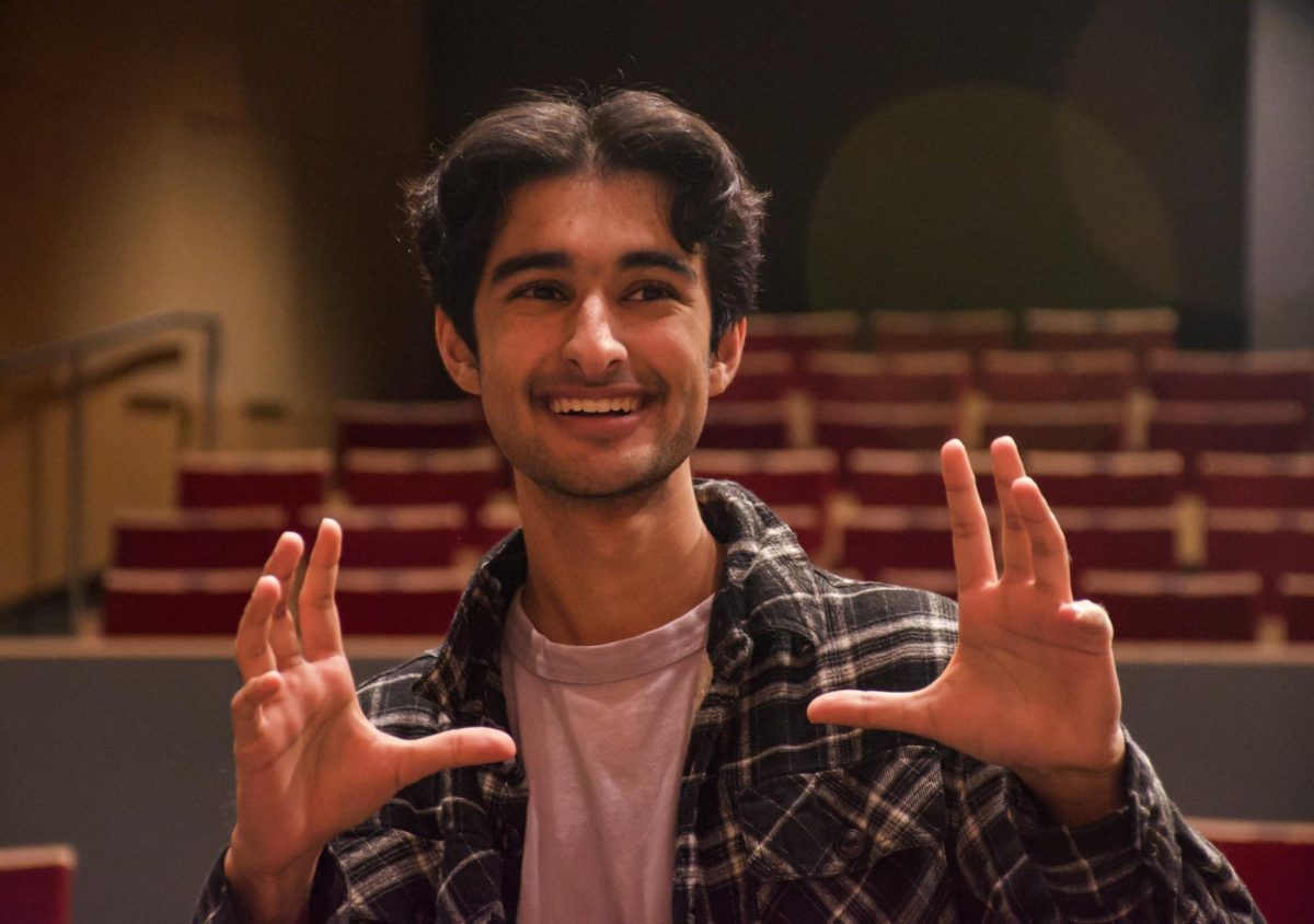
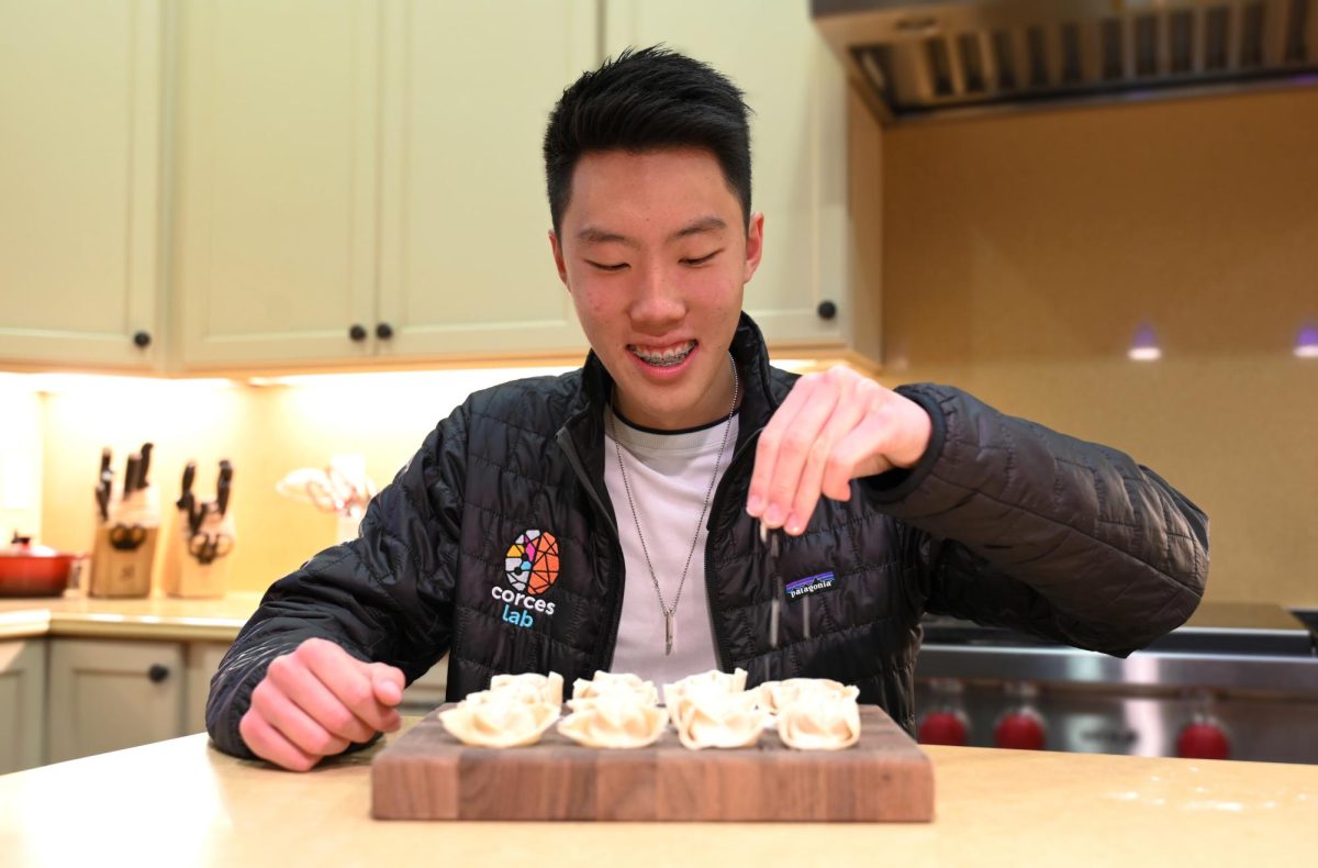
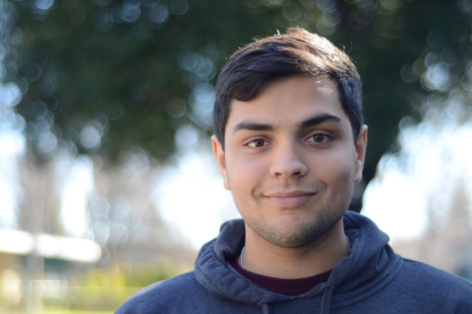
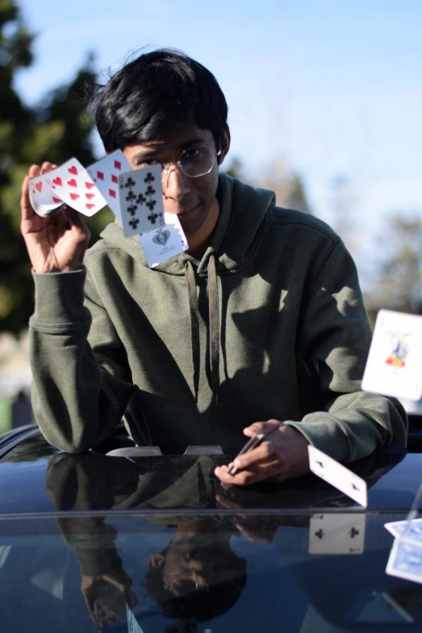
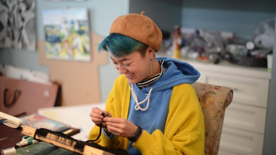
![“[Building nerf blasters] became this outlet of creativity for me that hasn't been matched by anything else. The process [of] making a build complete to your desire is such a painstakingly difficult process, but I've had to learn from [the skills needed from] soldering to proper painting. There's so many different options for everything, if you think about it, it exists. The best part is [that] if it doesn't exist, you can build it yourself," Ishaan Parate said.](https://harkeraquila.com/wp-content/uploads/2022/08/DSC_8149-900x604.jpg)
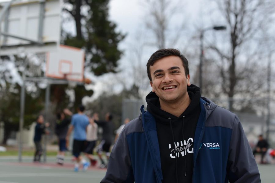
![“When I came into high school, I was ready to be a follower. But DECA was a game changer for me. It helped me overcome my fear of public speaking, and it's played such a major role in who I've become today. To be able to successfully lead a chapter of 150 students, an officer team and be one of the upperclassmen I once really admired is something I'm [really] proud of,” Anvitha Tummala ('21) said.](https://harkeraquila.com/wp-content/uploads/2021/07/Screen-Shot-2021-07-25-at-9.50.05-AM-900x594.png)
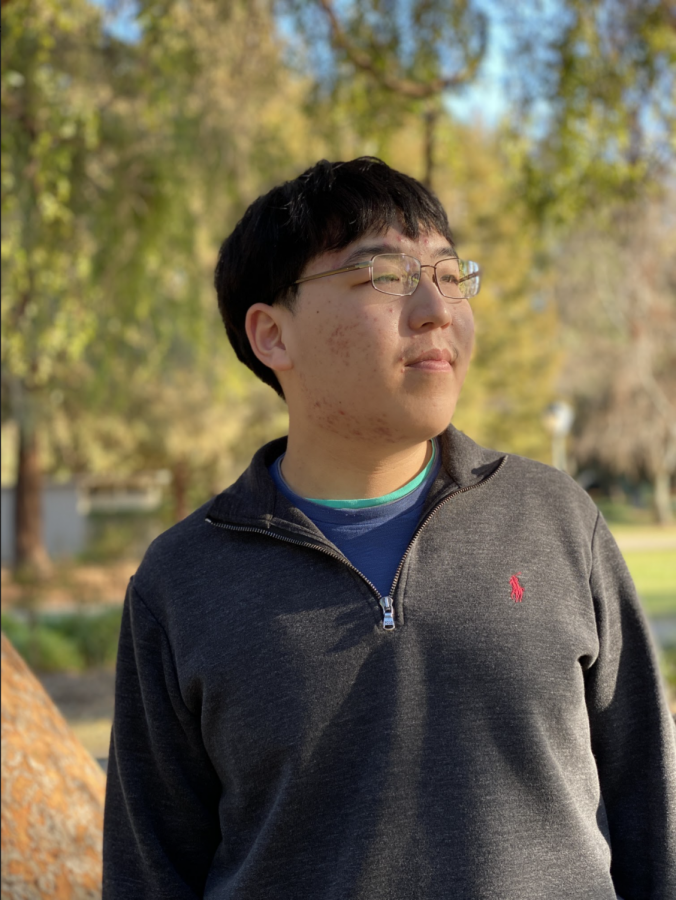


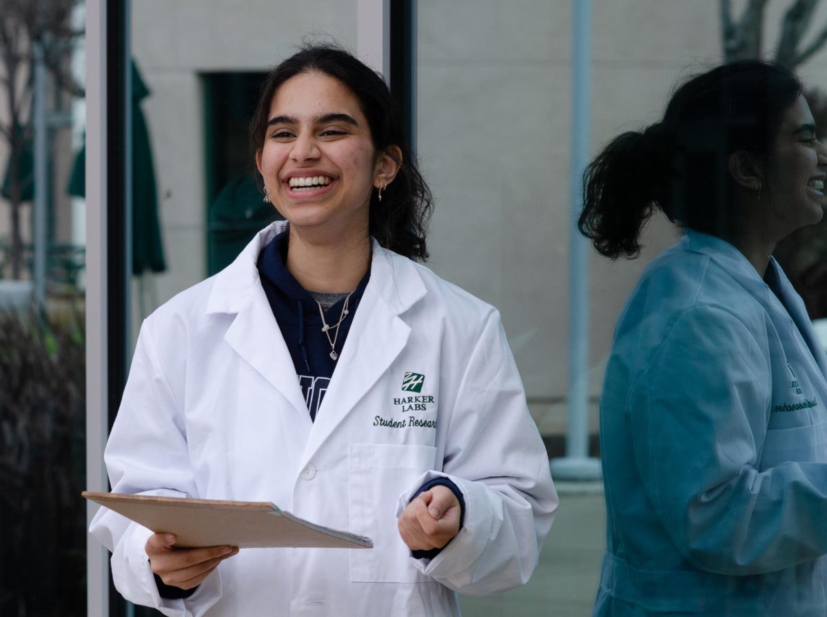
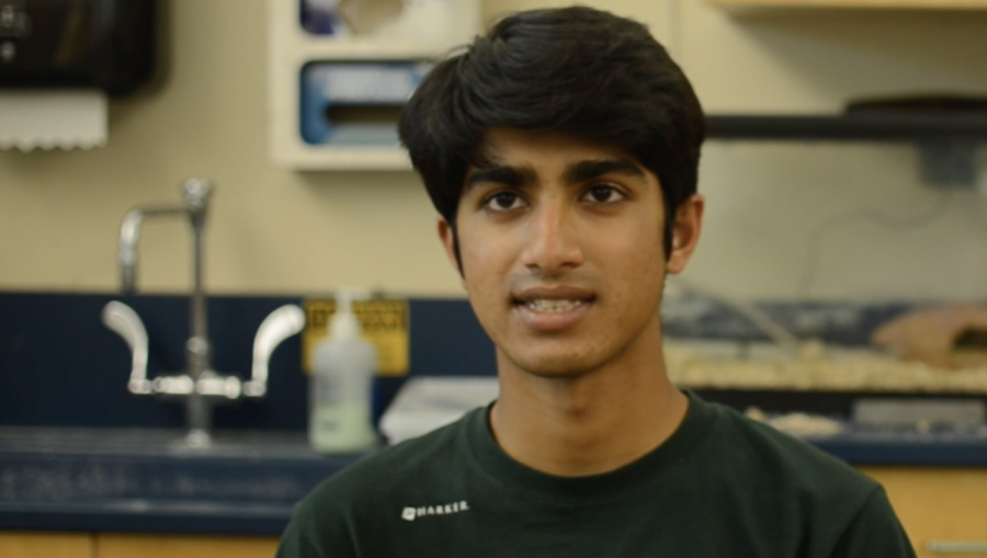
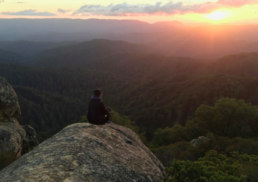
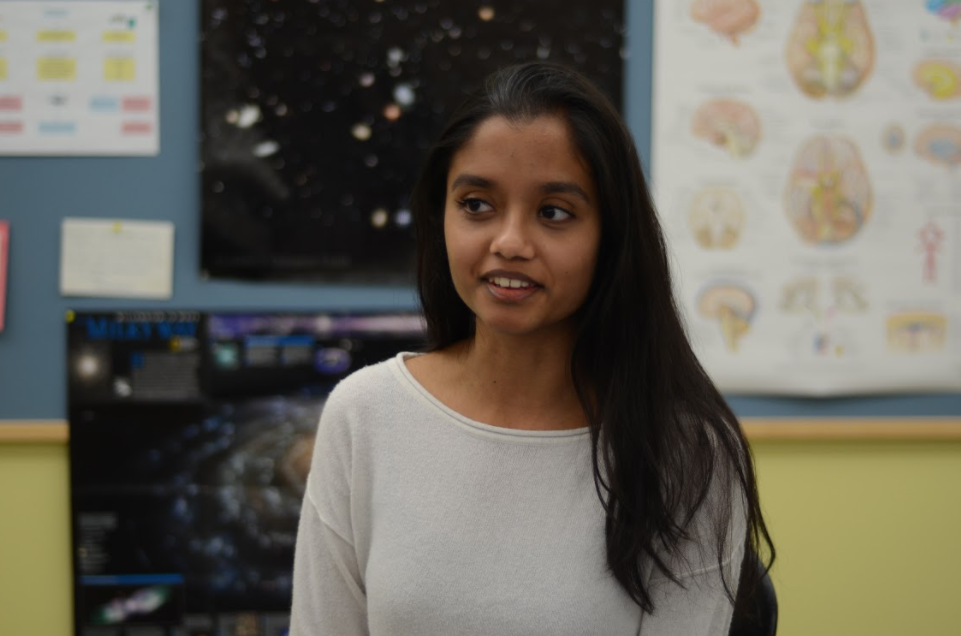
![“I think getting up in the morning and having a sense of purpose [is exciting]. I think without a certain amount of drive, life is kind of obsolete and mundane, and I think having that every single day is what makes each day unique and kind of makes life exciting,” Neymika Jain (12) said.](https://harkeraquila.com/wp-content/uploads/2017/06/Screen-Shot-2017-06-03-at-4.54.16-PM.png)

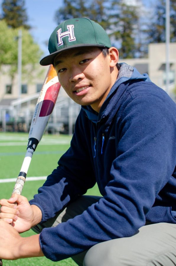
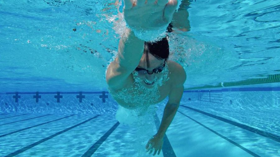
![“My slogan is ‘slow feet, don’t eat, and I’m hungry.’ You need to run fast to get where you are–you aren't going to get those championships if you aren't fast,” Angel Cervantes (12) said. “I want to do well in school on my tests and in track and win championships for my team. I live by that, [and] I can do that anywhere: in the classroom or on the field.”](https://harkeraquila.com/wp-content/uploads/2018/06/DSC5146-900x601.jpg)
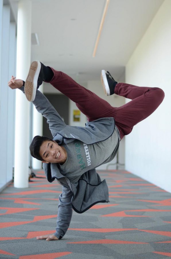
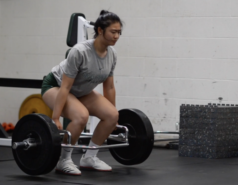
![“[Volleyball has] taught me how to fall correctly, and another thing it taught is that you don’t have to be the best at something to be good at it. If you just hit the ball in a smart way, then it still scores points and you’re good at it. You could be a background player and still make a much bigger impact on the team than you would think,” Anya Gert (’20) said.](https://harkeraquila.com/wp-content/uploads/2020/06/AnnaGert_JinTuan_HoHPhotoEdited-600x900.jpeg)
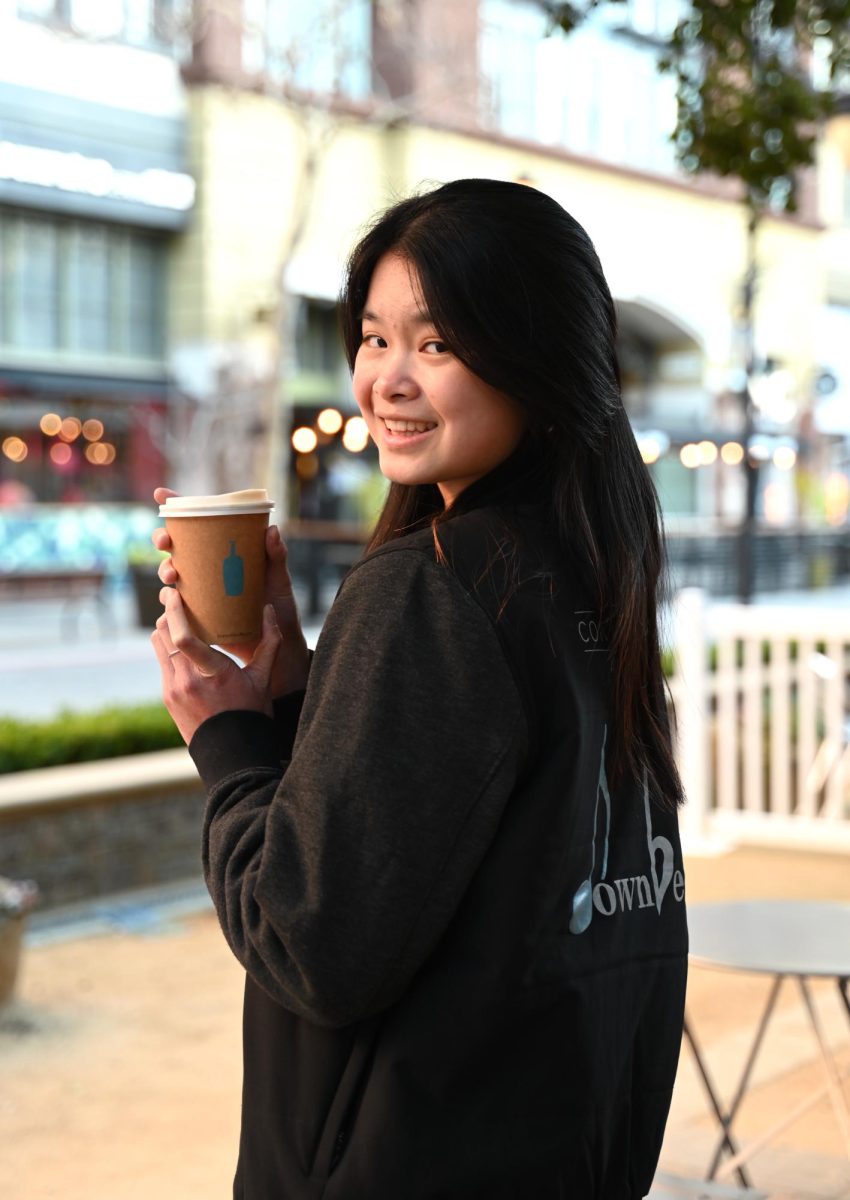
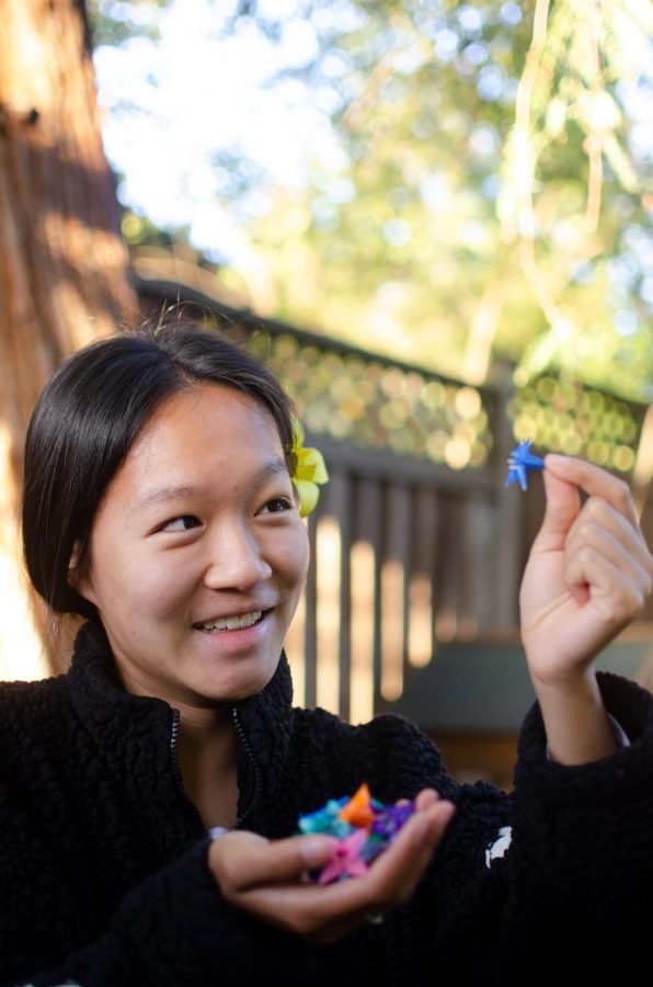
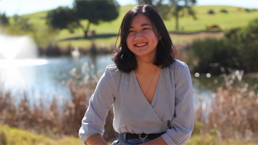
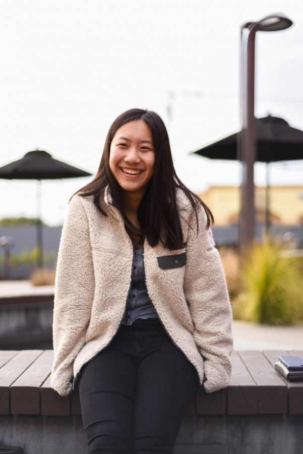
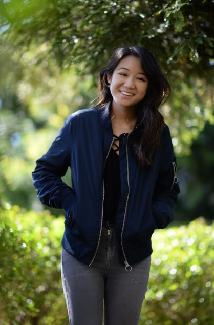
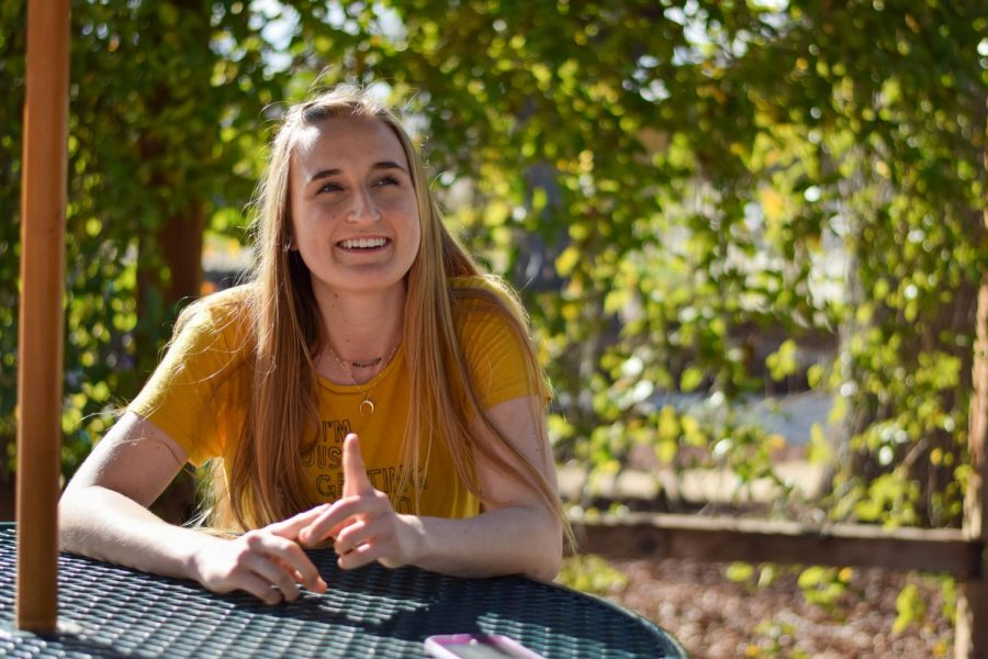
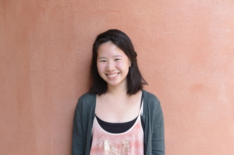
![“I'm not nearly there yet, but [my confidence has] definitely been getting better since I was pretty shy and timid coming into Harker my freshman year. I know that there's a lot of people that are really confident in what they do, and I really admire them. Everyone's so driven and that has really pushed me to kind of try to find my own place in high school and be more confident,” Alyssa Huang (’20) said.](https://harkeraquila.com/wp-content/uploads/2020/06/AlyssaHuang_EmilyChen_HoHPhoto-900x749.jpeg)
Cutting edges: New virtual dissection table
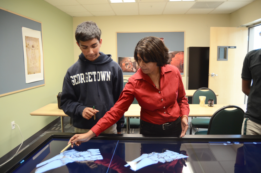
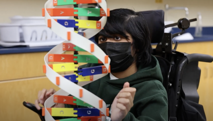
Human Anatomy and Physiology (HAPy) teacher Anita Chetty points out an ankle fracture to Ayush Midha (12). Currently, HAPy and some biology students can access the table to see structures in cadavers.
With a press of a button, a student takes a midline cut of a man. Inside, his organs reveal themselves, along with the violent purple cancer spreading through his intestines. With a slide of a lever, the purple is quickly replaced by a healthy pink color.
This scene, however, is not performed on a real man, but on a virtual touchscreen machine. The Upper School recently became the first high school in the world to buy an Anatomage 3D interactive virtual cadaver dissection table.
Prior to attaining the 3D interactive virtual cadaver dissection table, students would only have been able to learn through 2D figures such as models and pictures. The table allows students to virtually cut organs in different orientations and rotate them. Students have the opportunity to look inside a human body and go to any depth they want because of the images made by Computerized Tomography (CT) scans.
The table looks at real structures on human bodies, a feat which would only be possible if students had a physical cadaver to dissect. The table will primarily be used by Human Anatomy and Physiology (HAPy) students, but Biology students also have the opportunity to use the table during the human systems unit.
“The anatomy table takes us to another level in terms of understanding human anatomy and physiology,” Science Department Chair Anita Chetty said.
Students will have the opportunity to study human organs in detail and see their relationship to each other, as well as the pathology of organs when they begin to fail.
It shows a contrast between fully functioning and ordinary organs as well as abnormally functioning organs.
Pre-programed in the machine is a male cadaver who developed a metastatic melanoma and colon cancer. By looking through layers of the body, students can see how cancer spreads by examining which organs were the most affected to where the cancer eventually stopped spreading.
Not only are there pre-installed cuts, but students also have the ability to slice through section of the body by moving a slider and pressing a button. Once the virtual cut is made, the student can shift the organs around but cannot virtually take them out. Use of the virtual table avoids a process which has a higher risk of error – using a scalpel to physically cut through muscle and skin.
“[The table] was super cool because it was a lot bigger than I thought it would be; it was close to life size and really high definition,” HAPy student Archana Podury (12) said. “[The cadavers were those of] real people, and she told us about their stories. Most were really young people who died. One patient was a guy from Korea. His parents wouldn’t let him donate his body, [so the makers] got a graphic designer to redesign his face. We paid respect to the people who donated their organs [before we used the table].”
The table was acquired as a result of six parents who each donated $10,000.
“In terms of impact, it’s just incredible that this small group of parents are actually impacting the lives of so many other people,” Chetty said.
This article was originally published in the pages of The Winged Post on Oct. 17, 2014.
Priscilla Pan is the features editor for the Winged Post and co-creator of In a Nutshell. She is a senior and has been part of the journalism program for...