


![“[Building nerf blasters] became this outlet of creativity for me that hasn't been matched by anything else. The process [of] making a build complete to your desire is such a painstakingly difficult process, but I've had to learn from [the skills needed from] soldering to proper painting. There's so many different options for everything, if you think about it, it exists. The best part is [that] if it doesn't exist, you can build it yourself," Ishaan Parate said.](https://harkeraquila.com/wp-content/uploads/2022/08/DSC_8149-900x604.jpg)
![“When I came into high school, I was ready to be a follower. But DECA was a game changer for me. It helped me overcome my fear of public speaking, and it's played such a major role in who I've become today. To be able to successfully lead a chapter of 150 students, an officer team and be one of the upperclassmen I once really admired is something I'm [really] proud of,” Anvitha Tummala ('21) said.](https://harkeraquila.com/wp-content/uploads/2021/07/Screen-Shot-2021-07-25-at-9.50.05-AM-900x594.png)


![“I think getting up in the morning and having a sense of purpose [is exciting]. I think without a certain amount of drive, life is kind of obsolete and mundane, and I think having that every single day is what makes each day unique and kind of makes life exciting,” Neymika Jain (12) said.](https://harkeraquila.com/wp-content/uploads/2017/06/Screen-Shot-2017-06-03-at-4.54.16-PM.png)


![“My slogan is ‘slow feet, don’t eat, and I’m hungry.’ You need to run fast to get where you are–you aren't going to get those championships if you aren't fast,” Angel Cervantes (12) said. “I want to do well in school on my tests and in track and win championships for my team. I live by that, [and] I can do that anywhere: in the classroom or on the field.”](https://harkeraquila.com/wp-content/uploads/2018/06/DSC5146-900x601.jpg)
![“[Volleyball has] taught me how to fall correctly, and another thing it taught is that you don’t have to be the best at something to be good at it. If you just hit the ball in a smart way, then it still scores points and you’re good at it. You could be a background player and still make a much bigger impact on the team than you would think,” Anya Gert (’20) said.](https://harkeraquila.com/wp-content/uploads/2020/06/AnnaGert_JinTuan_HoHPhotoEdited-600x900.jpeg)

![“I'm not nearly there yet, but [my confidence has] definitely been getting better since I was pretty shy and timid coming into Harker my freshman year. I know that there's a lot of people that are really confident in what they do, and I really admire them. Everyone's so driven and that has really pushed me to kind of try to find my own place in high school and be more confident,” Alyssa Huang (’20) said.](https://harkeraquila.com/wp-content/uploads/2020/06/AlyssaHuang_EmilyChen_HoHPhoto-900x749.jpeg)
New 3-D virtual dissection table enriches Harker science classes
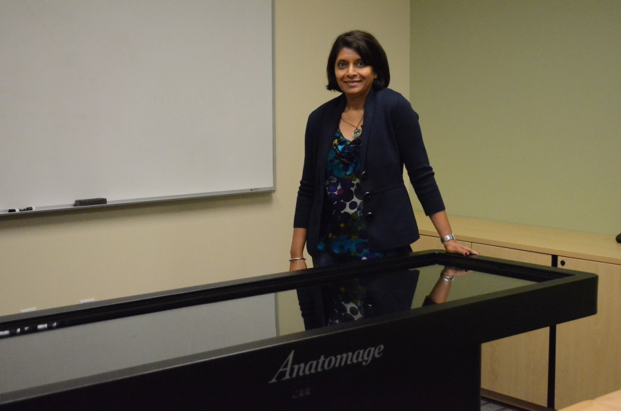
Harker acquired a technologically advanced visual anatomy table that will allow students to conduct virtual dissections. Anita Chetty is using the technology to enhance Harker’s science department and specifically, her Human Anatomy and Physiology classes.
Harker recently became the first high school in the world to acquire an advanced 3-D touch-interactive virtual cadaver dissection table.
The table is to be used primarily by the Human Anatomy and Physiology (HAPY) classes, but is also open to Biology students to view real dissections of preserved human and animal specimens.
“The main goal of this is to enhance the course and make the learning easier,” Science Department chair Anita Chetty said. “My whole philosophy about education is to have real life experiences. I am always looking for ways to make us more cutting edge.”
Using touch screen technology, digital cadavers can be rotated, deskinned, and dissected in any direction among other features. The human cadavers are donated bodies that have been frozen, dissected, and then scanned using CT imaging. The images are then loaded into the machine to allow students to virtually dissect the bodies. The software has three preloaded human cadavers, conjoined twins, and various animal cadavers such as a turtle, mouse, and cat, all life-size and anatomically accurate. Graphic artists then add in a face and features that may have been altered during the freezing process, such as blood vessels.
Two years ago, Chetty first saw the virtual dissection table at Stanford Medical school. She invited Anatomage, the Bay Area based company that manufactures the tables, to the 2012 Science Research Symposium, where the technology drew the attention of Harker administrators Joe Rosenthal and Butch Keller, head of the Upper school.
Keller, a biology major in college, shared his excitement about the table.
“When I first saw that exhibited at the symposium a couple years ago, I didn’t literally run, but I went directly to Ms. Chetty and said how do we get this,” Keller said. “I really think it’s the coolest thing ever.”
The $60,000 product was funded by six parent donations. Chetty expressed gratitude for their contributions.
“Many of the parents who donated are in computer science and technology fields, and they really committed to advancing our program to another level,” she said. “The parents that donated money opened the eyes of the community to the possibilities [that] Computer Science has in application along with biology. I am so grateful to those parents.”
The concept for the table was initially developed at the Stanford Anatomy Lab, and Harker has the most up-to-date version of the technology. Anatomage incorporates a software that can convert 2-D images into 3 dimensions known as Invivo 5. The touch-screen table runs on a Windows operating system with a split screen that allows teachers to display half the cadaver one on side, and a powerpoint on the other. All the cadavers can be labeled, unlabeled, and annotated in any way for class activities or quizzes.
Human Anatomy and Physiology student Ashir Bansal (12) looks forward to using the new table.
“I think it’s a privilege like no other, we get to experience dissection on a level above that of the human cadaver,” Ashir said. “The digitization of the body of a real person allows us to review anatomy in a way that typically only medical school students can.”
The table is housed in a small specialized Human Anatomy classroom on the second floor of Nichols.
Megy Appalaraju (12) is the Sports Editor of Harker Aquila. This is her second year in the Harker journalism program. Her favorite part about journalism...