

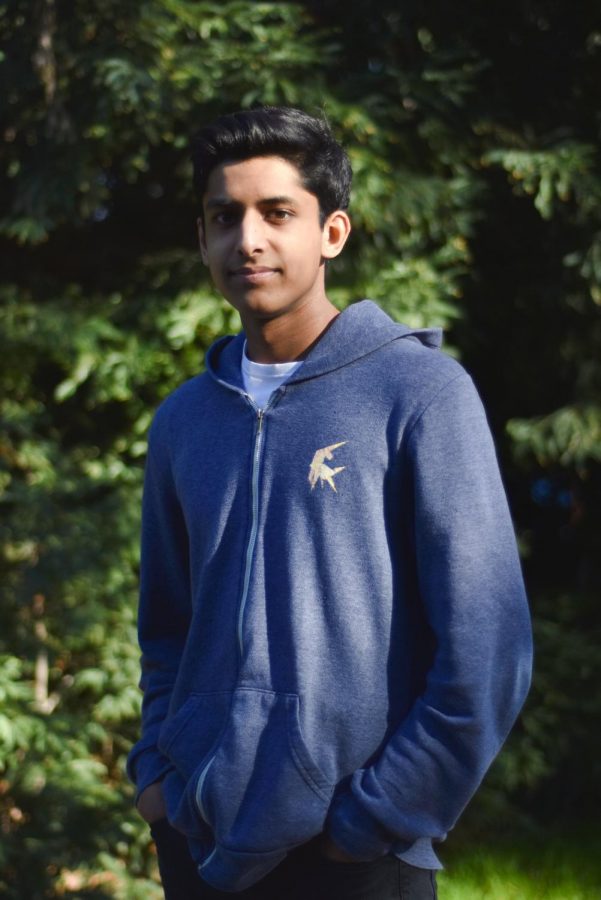
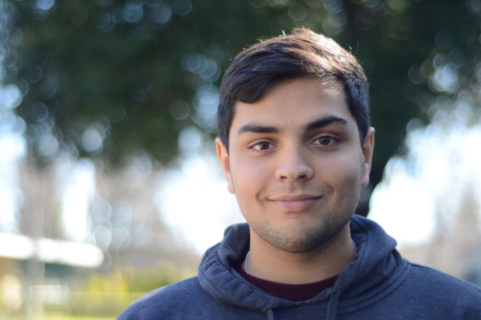
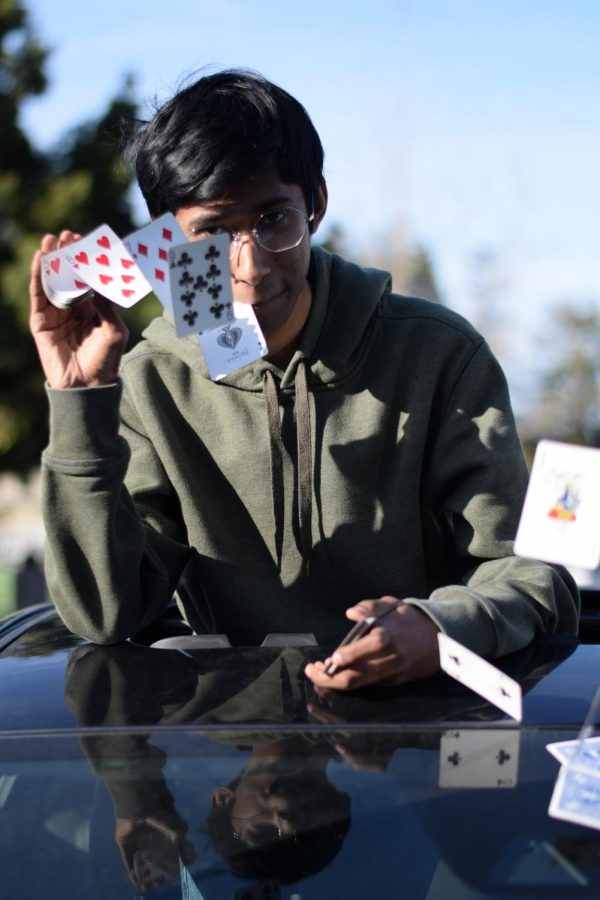
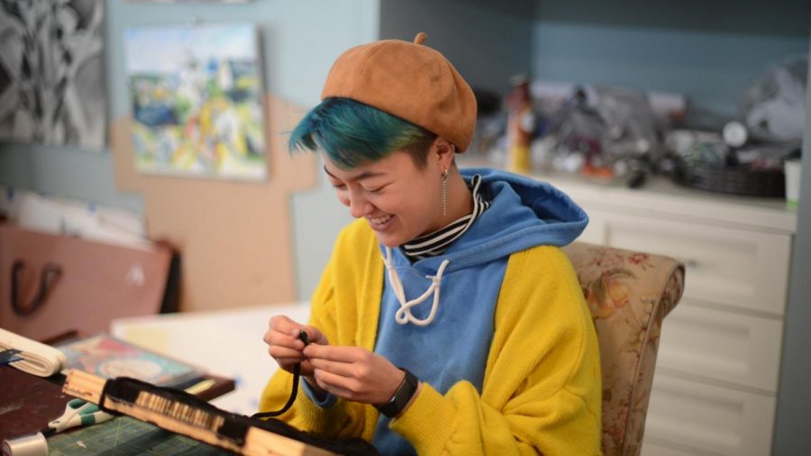
![“[Building nerf blasters] became this outlet of creativity for me that hasn't been matched by anything else. The process [of] making a build complete to your desire is such a painstakingly difficult process, but I've had to learn from [the skills needed from] soldering to proper painting. There's so many different options for everything, if you think about it, it exists. The best part is [that] if it doesn't exist, you can build it yourself," Ishaan Parate said.](https://harkeraquila.com/wp-content/uploads/2022/08/DSC_8149-900x604.jpg)
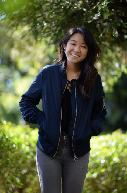
![“When I came into high school, I was ready to be a follower. But DECA was a game changer for me. It helped me overcome my fear of public speaking, and it's played such a major role in who I've become today. To be able to successfully lead a chapter of 150 students, an officer team and be one of the upperclassmen I once really admired is something I'm [really] proud of,” Anvitha Tummala ('21) said.](https://harkeraquila.com/wp-content/uploads/2021/07/Screen-Shot-2021-07-25-at-9.50.05-AM-900x594.png)
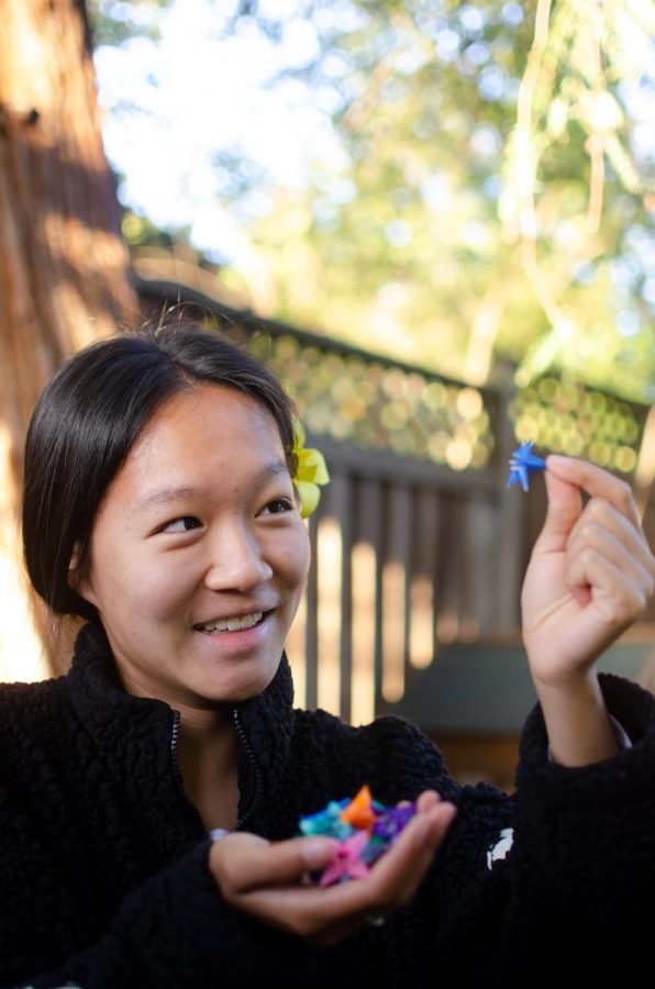
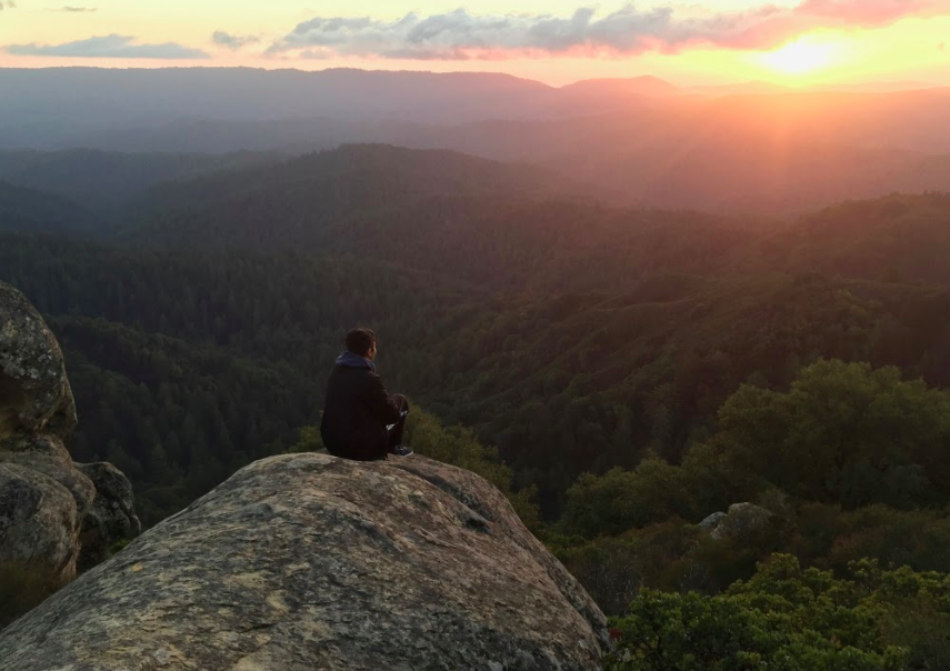
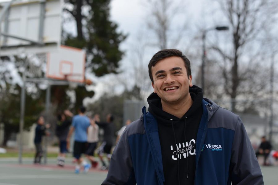
![“I think getting up in the morning and having a sense of purpose [is exciting]. I think without a certain amount of drive, life is kind of obsolete and mundane, and I think having that every single day is what makes each day unique and kind of makes life exciting,” Neymika Jain (12) said.](https://harkeraquila.com/wp-content/uploads/2017/06/Screen-Shot-2017-06-03-at-4.54.16-PM.png)

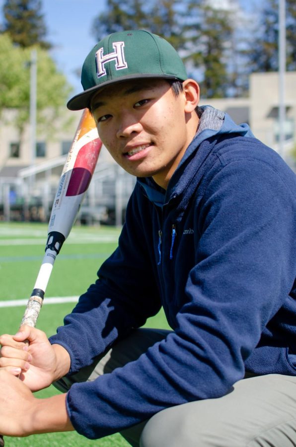
![“My slogan is ‘slow feet, don’t eat, and I’m hungry.’ You need to run fast to get where you are–you aren't going to get those championships if you aren't fast,” Angel Cervantes (12) said. “I want to do well in school on my tests and in track and win championships for my team. I live by that, [and] I can do that anywhere: in the classroom or on the field.”](https://harkeraquila.com/wp-content/uploads/2018/06/DSC5146-900x601.jpg)
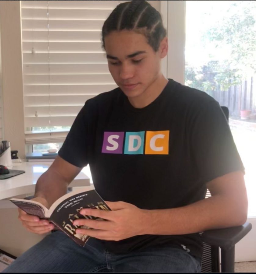
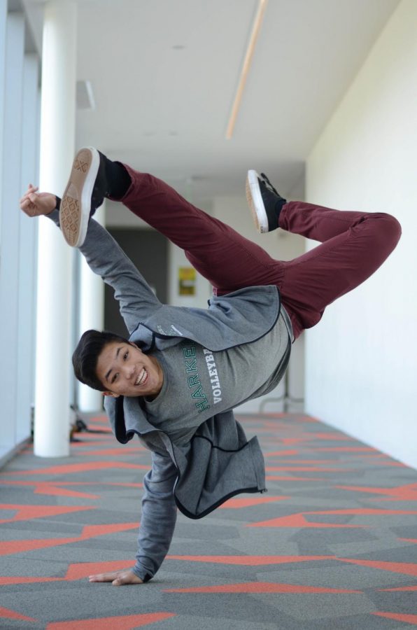
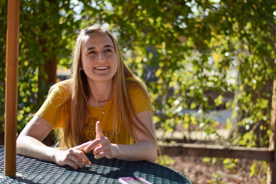
![“[Volleyball has] taught me how to fall correctly, and another thing it taught is that you don’t have to be the best at something to be good at it. If you just hit the ball in a smart way, then it still scores points and you’re good at it. You could be a background player and still make a much bigger impact on the team than you would think,” Anya Gert (’20) said.](https://harkeraquila.com/wp-content/uploads/2020/06/AnnaGert_JinTuan_HoHPhotoEdited-600x900.jpeg)
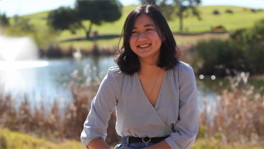
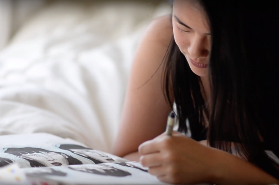
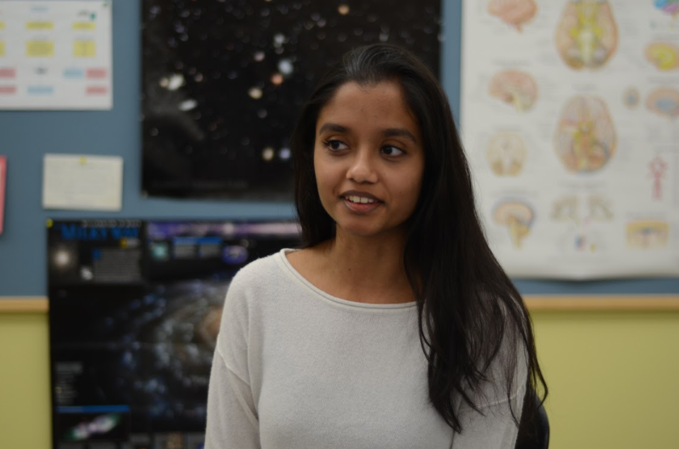
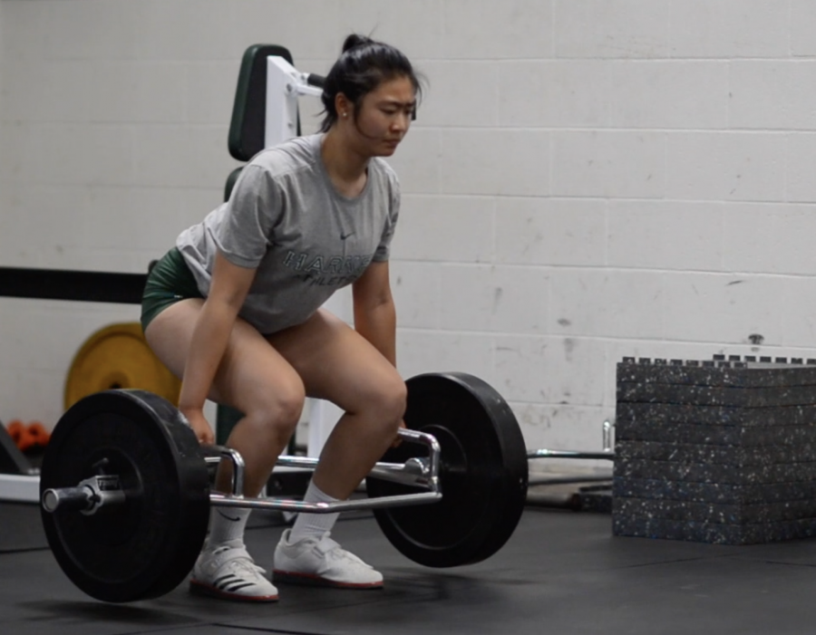
![“I'm not nearly there yet, but [my confidence has] definitely been getting better since I was pretty shy and timid coming into Harker my freshman year. I know that there's a lot of people that are really confident in what they do, and I really admire them. Everyone's so driven and that has really pushed me to kind of try to find my own place in high school and be more confident,” Alyssa Huang (’20) said.](https://harkeraquila.com/wp-content/uploads/2020/06/AlyssaHuang_EmilyChen_HoHPhoto-900x749.jpeg)
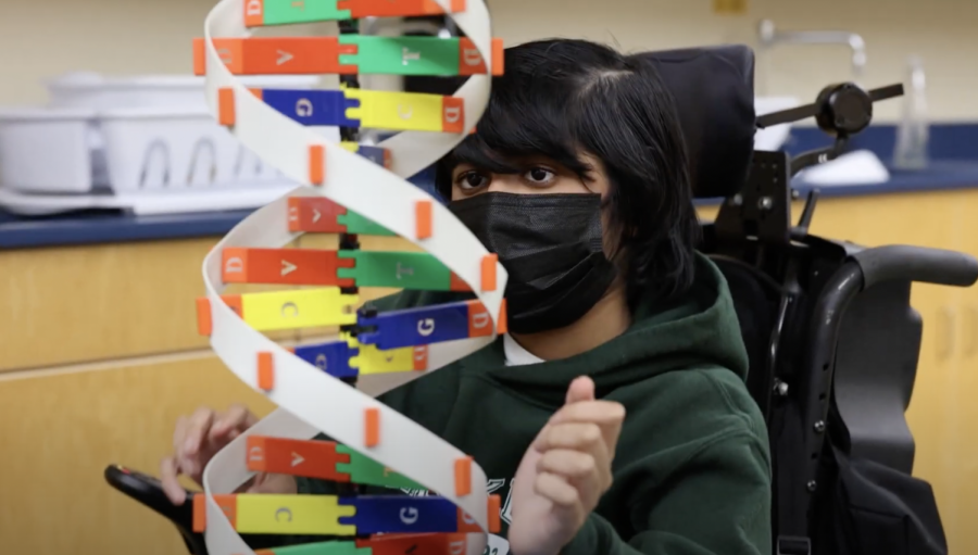
Human Anatomy students prepare microscopic slides in histopathology lab
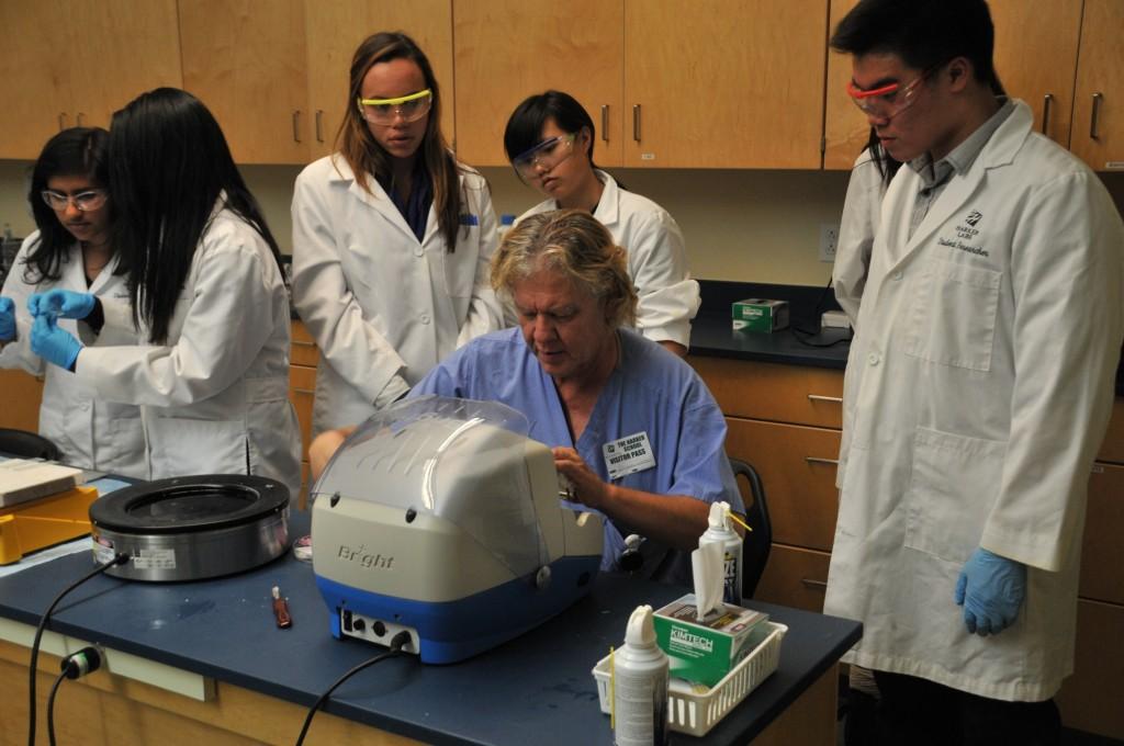
Histotechnician Chris Gallegos shows Human Anatomy and Physiology students how to operate a microtome to slice tissue samples. Students had the opportunity to prepare and view their own microscope slides, a process which involved cutting and staining the sample.
Human Anatomy and Physiology (HAPy) students experienced a slice of life as histologists, studying tissues during a lab led by professional histotechnician Chris Gallegos on Monday.
Gallegos works as a histotechnician at El Camino Hospital and also conducts his own research lab. He has been coming to HAPy classes for three years now to help with this histology lab.
Histology, or the study of tissues, is a topic most students have never seen before taking a human anatomy class. After studying various types of tissues in class, students had the opportunity to prepare their own samples to view under a microscope.
Students received a human tissue sample from a patient and had to prepare their own microscopic slide from that sample. This involved correctly cutting the tissue using a microtome, staining it through an extensive process, and properly mounting the sample onto a microscope slide.
“[Students] are so accustomed to being given the prepared slides,” HAPy teacher Anita Chetty said. “I wanted them to understand the process that’s involved in actually visualizing cells.”
In addition to preparing their own slides, Gallegos also brought a few gross anatomy samples: a portion of a cancerous human intestine and a human heart, both taken from autopsies, for students to look at and touch.
“It was kind of gross at first, but after a while, it was pretty cool,” Shivani Gillon (12) said. “The coolest part was definitely getting to hold a cancerous intestine.”
Chetty started the lab three years ago to further her philosophy of giving students a clinical experience in the field of human anatomy. After every unit, students complete some kind of clinical application component related to the organ system they are studying.
“I want it to be a real-life experience,” Chetty said. “I try to do that with many of my initiatives. We practice, we pretend, or we do the real thing, but in every case, it’s about ‘What would it be like to be in this actual situation?’”
A few of the other hands-on opportunities Chetty has planned for the rest of the year include looking at skin cancer with a recent PhD graduate after the integumentary unit of the class, and a cardiac surgery workshop in February where students will to insert a synthetic valve into a pig heart.